Tous droits réservés © NeurOreille (loi sur la propriété intellectuelle 85-660 du 3 juillet 1985). Ce produit ne peut être copié ou utilisé dans un but lucratif.
 Français
 English
 Español
 Português
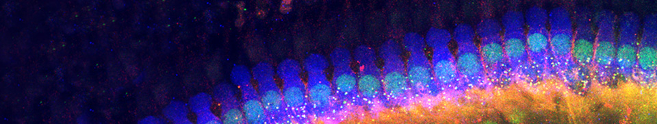
Inner hair cells (IHCs), the sensory cells of the cochlea, are responsible for signal transduction. Lying in a single row along the internal side of the tunnel of Corti, they are connected to type I spiral ganglion neurons (of which the axons represent about 95% of auditory nerve fibres). A human cochlea has between 3000 and 3500 IHCs.
Schematic of an inner hair cell (IHC)
")
The IHC and its innervation are schematically represented here on the left. Note, unlike in OHCs, the medial location of the nucleus, and the classic lateral plasma membrane.
Only a single synaptic complex is represented: i.e. a radial afferent bouton (blue) and a lateral efferent ending (pink). On average, one IHC is innervated by ten of these complexes.
IHC structure (TEM)
")
M Lenoir
IHC of a rat cochlea surrounded by its two support cells (with lighter cytoplasm), and rests on the inner pillar of the tunnel of Corti. It contains a central nucleus and its base is surrounded by the synaptic boutons of auditory nerve fibres’ afferent dendrites (dark grey), which can be seen entering the organ of Corti via the habenula perforata. |
")
R. Pujol, M. Vater IHC of a bat cochlea at the level of the ‘fovea’ (the area that codes the echo frequency) where up to 50 synaptic boutons can be seen for a single IHC (a)! |
Synaptic complex at the basal pole of an IHC

R Pujol |
This "synaptic complex" is composed of:
Scale bar: 350 nm |
 et la fibre afférente (a)")
R Pujol |
Typically, the synapse between IHC and auditory nerve endings is characterised by the presynaptic body surrounded by microvesicles (red arrow).
Pre- and postsynaptic densification of membranes is clearly visible (pink arrows), as well as an endocytotic profile (blue arrow) in the IHC. Scale bar: 200 nm |

R Pujol |
Synapse between a vesicular ending of the lateral efferent system, and an afferent dendrite beneath an IHC. The asterisk (yellow) indicates the post-synaptic density in the afferent fibre. Scale bar: 100 nm |
 qui vient de quitter la CCI")
R Pujol |
An afferent dendrite (a) leaving an IHC synapses with 3 vesiculated efferent endings (asterisks). This image is characteristic of the importance of the efferent control exerted on the auditory nerve fibres. The efferent neurotransmitter dopamine has been implicated in protection against glutamate excitotoxicity, and this is dicussed elsewhere (see " Physiology"). |
Facebook Twitter Google+