Tous droits réservés © NeurOreille (loi sur la propriété intellectuelle 85-660 du 3 juillet 1985). Ce produit ne peut être copié ou utilisé dans un but lucratif.
 Français
 English
 Español
 Português
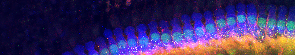
The organ of Corti, named after Alfonso Corti who first described it, is the sensorineural organ of the cochlea. It is composed of sensory cells called hair cells, nerve fibers that connect to them, and supporting structures.
Overview structure-function
Schematic of the organ of Corti

S. Blatrix |
1-Inner hair cell (IHC) |
In this transverse section of the basal part of a mammalian cochlea, 1 IHC (1) and 3 OHCs (2) are represented on either side of the tunnel of Corti (3). The tectorial membrane (6), floating in endolymph, caps the tallest stereocilia of the hair cells. The IHC is surrounded by supporting cells, whereas OHCs are anchored to the Deiters’ cells (7), their lateral membrane in direct contact with a fluid called corticolymph, similar to perilymph, which fills the tunnel of Corti (3) and Nuel’s spaces (8).
The cuticular plate of the hair cells, together with the head of the pillars, the phalangeal processes of Deiters’ cells and other cells such as Hensen’s cells (9) form the reticular lamina which seals the endolymphatic compartment. Nerve fibers arrive into or leave the organ of Corti through the basilar membrane (4), via the habenula perforata (5).

R. Pujol |
Transverse sections through guinea pig organ of Corti (Nomarski optics) See schematic above for the legends |
")
Functional scheme of the Organ of Corti
Animation: S. Blatrix, scientific concept G. Rebillard et R. Pujol
The function of the organ of Corti, for a soft sound (such as speech), can schematically be summed up in 5 sequences:
(1) Sound waves, transmitted by the perilymph, make the basilar membrane vibrate up and down. Passive tonotopy mobilises the basilar membrane from the base (high sounds) to the apex (low sounds) of the cochlea
(2) Stereocilia of the OHCs, embedded to the tectorial membrane, bend when the basilar membrane rises, causing the OHCs to depolarise (by the influx of K+ ions).
(3) Excited (depolarised) OHCs react by contracting (= electromotility). Due to the close link between the OHCs, the basilar membrane, and the reticular lamina, this active mechanism creates energy that amplifies the initial vibration. It also plays a role in active frequency filtering (active tonotopy).
(4) The IHC is excited, probably via direct contact with Hensen’s stripe within the tectorial membrane.
(5) The synapse between the IHC and the auditory nerve fibre is activated, and a message is sent to the brain.
Facebook Twitter Google+